Alo Yeditepe
Alo Yeditepe
Hip Impingement Syndrome
Yeditepe University Hospitals Orthopedics and Traumatology Specialist Prof. Dr. Gökhan Meriç answered questions about Hip Impingement Syndrome.
Hip Impingement Syndrome
The hip joint is the joint where your femur meets the pelvis. The hip bones are in the form of a round knob and a nest in which it sits. Normally, this joint moves smoothly during walking, running, sitting, and getting up. Hip impingement syndrome is caused by a bone protrusion in the hip joint or damage to the soft tissues in this area, causing difficulty in movement of the joint and pain.
Conditions that increase the risk of hip compression; genetic characteristics of the person, recurrent severe hip exercise in athletes, previous hip trauma, and those with childhood hip disease are more likely to develop.
Hip impingement syndrome can be seen as an over-wrapping of a protrusion in the hip knob bone or the bone surrounding the hip joint, or as these two conditions are together. The hip bone protrusion is more common in young men, and excessive clamping of the hip joint is more common in women. It may cause symptoms by causing damage to the labrum, the soft tissue surrounding the articular cartilage, and the hip joint due to compression during movement.
What are the Symptoms of Hip Impingement?
Patients may experience hip impingement for years, and you may not know it because it is usually not painful in its early stages. Symptoms of hip compression can be seen as "pain" in the groin and limitation and strain in the movements of the hip, especially when walking or flexing the hip. It can occur during movements such as getting out of the car, riding a bicycle, or tying your shoes.
Weakness in the hip muscles, and repetitive strain may cause complaints of people who are prone to hip compression syndrome. Weakness of the hip muscles increases the joint load by shifting the hip bone forward. Damage to the capsule around the hip joint may cause tears.
Who Can Have Hip Impingement Syndrome?
- People with a family history of hip impingement syndrome
- Repeated stress during childhood and adolescence in people who engage in repetitive activities during development, such as basketball or football, that often require intense hip movement, can trigger hip bone remodeling, and eventually bone development that creates compression.
- History of childhood hip disease that may have changed the shape of the hip bone head (e.g., hip dislocation, slipped femoral head epiphysis, or Perthes disease)
- History of hip fracture
Does Hip Impingement Syndrome Cause Osteoarthritis?
There is a close relationship between hip compression and hip arthritis, but it does not develop arthritis in everyone who experiences it. When the ball and the seat of the hip are not fully seated together, the articular cartilage covering their surfaces can be subjected to excessive mechanical friction. This friction erodes the cartilage. In some cases, the hip cartilage may partially detach from the bone, causing joint cartilage damage and hip osteoarthritis.
What Kind of Symptoms Does Hip Impingement Show?
- Significant hip or inguinal pain related to certain movements or positions, such as going up and down stairs
- Restriction of movement in the hip joint
- Clicking and noise in the hip joint
- Pain during bending, tying shoes and getting out of the car, and getting on in daily life
- Pain in sports activities that strain the hip, such as squats and deadlifts
What Activities and Exercises Should People with Hip Impingement Syndrome Avoid?
People with hip compression should avoid crouching, sitting still for a long time, and movements that require sudden hip rotation. Sudden turn and squat exercises should be avoided while exercising as they may increase the complaints of the patients.
Which Tests Are Evaluated in the Diagnosis of Hip Impingement Syndrome?
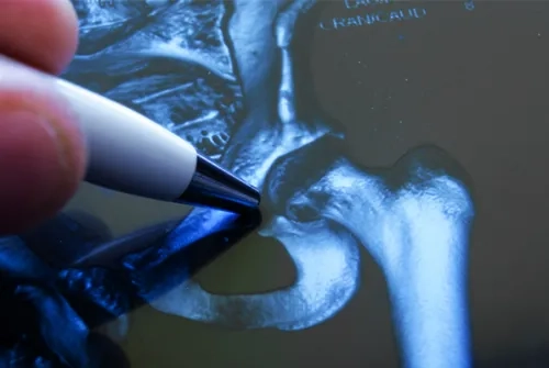
For the diagnosis of hip impingement syndrome, first of all, an examination of hip movements and some special tests are applied. Hip films are important in the evaluation of bone protrusions and compression types in imaging methods. With MR imaging, it is evaluated whether there is soft tissue damage that may accompany compression.
What are the Treatment Options for Hip Impingement Syndrome?
Treatment of hip impingement syndrome varies according to the degree of impingement and concomitant pathologies. In the initial period, exercises that strengthen the muscles around the painkillers hip are recommended. In cases where pain and limitation of movement persist, it is aimed to reduce edema and pain around the hip with physical therapy and injections containing corticosteroids to the hip joint. Despite all these treatments, hip arthroscopy is planned in case of advanced compression and accompanying capsule rupture, where complaints continue. It is a closed intervention performed by entering through 2-3 small holes in hip arthroscopy. In this process, the bone structures that cause compression are removed and the accompanying capsular tear, if any, can be repaired. After the intervention, patients are allowed to stand up with crutches for a while. Physical therapy is given again after the intervention. Open surgeries may be required in patients with advanced cartilage abrasion in the hip joint due to compression where complaints are advanced.
This content was prepared by Yeditepe University Hospitals Medical Editorial Board.
”
See Also
- Robotic Hip Replacement Surgery
- Knuckle Cracking Can Be Dangerous When It Becomes a Habit!
- What is Hallux Rigidus (Stiff Big Toe/Toe Arthritis)? Symptoms and Treatment
- What is Hallux Valgus (Bunion)? How is it Treated?
- Meniscus and Cartilage Transplantation Can Be Done at the Same Time
- Ergonomics in Automobiles Prevents Accidents
- Joint Pain and Causes of Joint Pain
- Walking and Returning to Social Life After Knee Replacement Surgery
- Robotic Knee Prosthesis Surgery
- Patients Who Undergo Joint Replacement Surgery Can Stand Up and Walk the Next Day
- Big Toes Can Be A Big Problem!
- What is Synovitis in Joints?
- What is the Future of Treating Cartilage Problems?
- Not Only Athletes Suffer from Meniscus Tears!
- Is Cartilage Damage More Common in Those Who Run for Long Periods?
- Beware If Your Shoulder Or Neck Pain Lasts Longer Than Two Days After Swimming!
- Mistakes While Swimming Can Cause Shoulder Pain
- Myths About Fractures
- Vitamin D Deficiency May Be The Cause Of Your Joint Pain
- Have Your Baby Take Their First Steps in Good Health
- Heavy School Bags Can Cause Pain in the Lower Back, Shoulders, and Hands!
- Sports Injuries
- Cartilage Transplantation from Donor Has Been Started to Perform in Turkey
- Working From Home Increases Waist and Neck Problems
- First Cartilage Transplant from a Donor
- Pay Attention to the Pain That Occurs in the Front Part of Your Knees While Playing Sports!
- Young Patient Who Could Not Walk Due to Cartilage Damage Recovered With Cartilage Transplantation
- Knee Arthritis
- Knee Pain
- Crunching in the Kneecap May Be a Sign of Calcification
- Injured Athletes Can Return To Sports With Cartilage Transplant
Alo Yeditepe


