Alo Yeditepe
Alo Yeditepe
Lumbar Spinal Stenosis
What is Lumbar Spinal Stenosis?
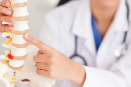
The bone structure that protects the spinal cord and nerve fibers is called "vertebrae." The spinal cord and nerve fibers descend along the canal at the center of the vertebrae from the nape of the neck to the coccyx. For the spinal cord and nerves to be present in this canal inside the vertebrae in an unconstraint manner, the canal's diameter should not be below 11 mm. Conditions in which the diameter is below this critical value are called ‘spinal stenosis’ or ‘narrowed canal.’
What Causes Narrowed Canal?
Deformations that develop due to aging are the most important cause of a narrowed canal. In general, the narrowed canal is most common among people with a congenitally narrow canal. Another cause of spinal stenosis is the growth of vertebrae due to overloads in the areas where they form joints.
What are the Symptoms?
Spinal stenosis, even if very severe, may not always give symptoms. Its findings develop insidiously gradually over time.
The symptoms are pain, numbness, and cramping in the lower back, back, or legs. In addition to weakness in the legs, it may cause bladder and/or intestinal problems in rare cases. Complaints increase with standing and walking for a long time. After walking for a limited time, the patient may need to stop and crouch down due to weakness and numbness in the legs. The walking distance may gradually decrease. The pain may be relieved or clear up by bending or sitting. The following quote exemplifies the typical complaint of the patients: "while I used to be able to walk comfortably for 2-3 km, now my walking distance has been restricted so much, I immediately feel the desire to sit down and rest."
How is the Diagnosis Made?
The degree of spinal stenosis can be determined in detail through magnetic resonance imaging (MRI) performed after the examination. Computerized tomography (CT) can be used to examine bone structures.
When deciding on treatment, whether the patient's complaints, endurance, and quality of life have decreased is assessed. For stenoses that cause pain, non-surgical options can be tried first. These are non-steroidal anti-inflammatory drugs (NSAIDs) and analgesics. Steroid and local anesthetic injections can also be performed directly to the facet joints in the spine or to the epidural space (the space between the membranes surrounding the nerves). Overusing drugs does not lead to faster recovery and may cause undesired drug side effects.
What are the Treatment Options?
Conservative (Non-surgical) treatment options other than drug therapy:
Physical therapy or an exercise program that strengthens the back, abdomen, and leg muscles is important for strengthening the muscles and restoring mobility. Aerobics, cycling, and walking are recommended. Because such actions increase the amount of blood coming to the nerves and thus reduce the symptoms of narrowing.
Spinal stenosis may not constitute a dangerous condition in adults unless significant and progressive leg weakness develops and there are bladder or intestinal problems. However, non-surgical methods cannot mechanically correct the stenosis in the spinal canal.
Surgical Treatment:
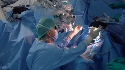
Surgical methods are preferred in patients whose pain does not subside through non-surgical methods, whose complaints increase over time, or who have progressive leg weakness and bladder and bowel problems. The surgical procedure aims to eliminate the pressure and expand the canal diameter (decompression). With surgical treatment, patients can regain their former walking capacity. Leg pains and weakness improve. Currently, patients can return to their normal lives a few weeks after surgery.
If lumbar spondylolisthesis accompanies narrowed canal or in very advanced narrowing, spinal fusion (vertebral fusion) surgery may also be required in addition to decompression. Titanium implants can be applied to the bone tissue between the vertebrae to be joined in this procedure.
This content was prepared by Yeditepe University Hospitals Medical Editorial Board.
”
See Also
- What is a Brain Tumor? Symptoms, Causes, and Treatment
- Causes and Treatment Methods of Cervical Herniated Disc
- What is Parkinson's Surgery?
- Brain / Spinal Cord Spine and Nerve Surgery (Neurosurgery)
- Lumbar Spondylolisthesis (Lumbar Slipped Vertebrae)
- Spinal Fractures
- Narrow Cervical Canal and Myelopathy
- Use of Vitamin D in Pregnant Women
- Brain Battery Connecting Parkinson Patients to Life
- Treatment Success in Brain Tumors Also Depends on the Family
- Not All Hand Tremors Are Caused by the Parkinson's Disease
- Do not Fill Your Brain With Junk Food If You Want A Good Memory
- When Does the Spine Require Surgical Treatment?
- Osteoporotic Spinal Fractures Can Cause Bigger Problems If Not Treated Early
- Patient Selection is Important in Parkinson's Surgery
- Canal Narrowing in the Spinal Cord
- Beware If Your Walking Distance Is Decreasing!
Alo Yeditepe


