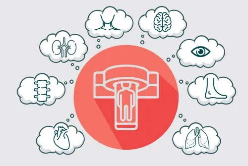
Patients are given very low amounts of targeted radioactive substances as food/drink, intravenously, by inhalation, or by injection into or under the skin. This given radioactive substance goes to the targeted organ or tissue and provides uptake there. These regions are then viewed with special cameras. The radiation received by the patient during this procedure is at a very low level, considering the benefit-harm balance. All these examinations are evaluated by Nuclear Medicine specialists.
Nuclear Medicine Technologies
PET (Positron Emission Tomography) / CT (Computed Tomography)
By using a small amount of radioactive substance, high-resolution and quality images are provided. Respiratory and cardiac-triggered PET/CT scans can also be performed. In this way, the amount of radiation that patients are exposed to is greatly reduced. Detailed examinations of the patients are done without unnecessary radiation.
SPECT (Single Photon Emission Tomography) / CT (Computed Tomography)
Other scintigraphic examinations are performed with this imaging system. Thanks to this system, besides the functional image obtained by scintigraphy, the anatomical image obtained by CT is superimposed, increasing the accuracy of diagnosis. Again, thanks to this system, dosimetric calculations, which can be applied in very few centers in our country, can be made with high accuracy, especially in the treatment of thyroid cancer, prostate cancer, and neuroendocrine tumors, and patients can be treated with personalized radioactive substance amounts.
Bone Mineral Densitometry (DEXA)
It is a device called dual-energy X-ray absorptiometry, which is used to measure bone mineral loss. Bone densitometry is often used in the diagnosis of osteoporosis, which affects postmenopausal women but can also be seen in men. In addition, DEXA is also effective in evaluating the response to treatment in osteoporosis and other conditions leading to bone loss.
Urea Breath Test
It is a simple and reliable test for detecting Helicobacter pylori, a bacterium that causes gastritis and peptic ulcer. Helicobacter pylori is a bacterium that multiplies by settling in the gastric or duodenal (first part of the small intestine) mucosa in humans and causes a wide range of stomach diseases ranging from gastritis to ulcers, from stomach cancer to gastric lymphoma. Making the correct diagnosis of Helicobacter pylori is the first step in ulcer treatment.
It also prevents more serious digestive system problems.
Thyroid Uptake Test
The thyroid uptake test is a method used to evaluate the function of the thyroid gland. It is used to determine the rate of radioiodine retained in the thyroid gland after oral ingestion of radioactive iodine. It provides guidance for determining the amount of I-131 to be given due to hyperthyroidism, making the differential diagnosis of diseases causing hyperthyroidism, and calculating the radioactive iodine treatment dose in patients who have undergone surgery for thyroid cancer.
Gamma Probe
The gamma probe is a pen-shaped probe that can detect low amounts of radioactive substances and is a medical device used for sentinel lymph node (SNL) sampling during surgery. The first lymph node to which many tumors, especially breast cancer and malignant melanoma can spread, is removed with the help of this device.
Before the operation, on the morning of the operation or the day before, the SLN is found with special methods by an experienced Nuclear Medicine physician in the Nuclear Medicine department, the lymph node is marked and the patient is directed to surgery. During surgery, the pre-marked SLN is removed by the surgeon with the help of Gamma Probe, and the material is sent to pathology to be frozen. If cancer cells are detected as a result of pathology, that area is completely cleaned. If cancer cells are not detected, unnecessary procedures are not performed on that area. Thus, the operation time is shortened, and postoperative complications are reduced.
Synthesis Unit
Some tumors, such as prostate or neuroendocrine tumors, do not take up the FDG used in the classical method, PET/CT. With the help of compounds marked with the Ga-68 radioactive substance, prostate cancer or neuroendocrine tumor imaging, which cannot be performed with conventional FDG PET imaging, can be performed. Again, with the help of this synthesis unit, it is possible to perform PET imaging for different purposes by using many newly developed radioactive compounds.
Treatments Applied in Nuclear Medicine
- “Iodine I-131” for the treatment of thyroid cancers
- “Lutetium Therapy” in Neuroendocrine Tumors
- “Lu-117 PSMA” for the treatment of advanced hormone-resistant prostate cancer
- “Ra-223” in prostate cancer patients with advanced hormone-resistant bone metastases
- “Gallium” in prostate cancer
- Radioembolization with “Y-90” microspheres in primary or metastatic liver tumors
- Palliative pain in patients with bone metastases
- Radionuclide for intra-articular fluid accumulation and pain
- “Iodine-131” for the treatment of hyperthyroidism (toxic goiter)
- Sentinel lymph node removal with intraoperative gamma probe in diseases such as breast, malignant melanoma, and parathyroid adenoma.


