Alo Yeditepe
Alo Yeditepe
Goiter (Thyroid Gland) Biopsy
What is Thyroid Gland?
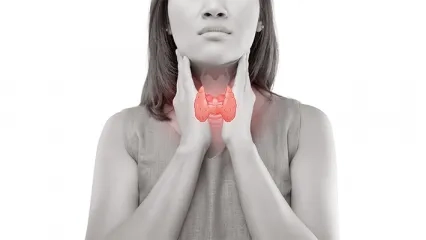
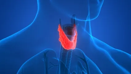
The thyroid gland is a butterfly-shaped organ that is located in the front part of the neck. Goiter, on the other hand, is a term for a disease that causes the thyroid gland to grow.
Enlarged thyroid gland (goiter disease) often occurs with an equal level of an enlarged gland on all sides. You can think of this situation as when air is blown into a butterfly-shaped balloon, and its whole side swells to the same level.
A less common condition is an enlargement of the thyroid gland by forming nodules. These bumps may vary in size from several millimeters to several centimeters.
The most important feature of tubers is the risk of transformation into a malignant disease (cancer). The most reliable way to understand this is to enter the lump with a needle and take some cells and examine these cells under a microscope. The thickness of the needle used during this procedure is not different from the needle used to draw blood or to inject medication from the hip.
During the procedure, we will ask you to lie on your back and remain in a stationary position.
In our university, every biopsy is performed with ultrasonographic imaging. After the location of the lump is determined by ultrasound, the cells are removed by sending the needle safely into the lump. The process is terminated within a few minutes.
Before each biopsy procedure, a regional anesthetic (pain-relieving) substance is applied at our university. Thus, by numbing that area, the pain is reduced to the lowest level.
The received cells are delivered to the pathology department in a special liquid. Depending on the intensity of the pathology department, your report will be out in a few days. You should receive your report and forward this examination to your physician who requests it from you.
How Does Thyroid Biopsy Work?
During the procedure, we will ask you to lie on your back and remain in a stationary position.
In our university, every biopsy is performed with ultrasonographic imaging. After the location of the lump is determined by ultrasound, the cells are removed by sending the needle safely into the lump. The process is terminated within a few minutes.
Before each biopsy procedure, a regional anesthetic (pain-relieving) substance is applied at our university. Thus, by numbing that area, the pain is reduced to the lowest level.
The received cells are delivered to the pathology department in a special liquid. Depending on the intensity of the pathology department, your report will be out in a few days. You should receive your report and forward this examination to your physician who requests it from you.
It is very important to pay attention to the following in order not to have problems during a goiter biopsy:
- Have a light breakfast in the morning. Do not come hungry.
- Do not take blood thinners or aspirin if you are using them. You need to stop these medicines at least 7 days in advance. If you have taken any of these medicines in the last 7 days, do not have a biopsy. Talk with your physician about the condition.
- If you take any other medicines besides blood thinners or aspirin regularly, you can take them on time and at your usual dose.
- Remove any necklace before the biopsy.
- Choose clothes that are open at the neck or that can be opened with buttons.
- Do not make a bun from the back if you have long hair. During the procedure, you will be asked to lie down with your head pushed back. The presence of the knob will disturb you in this position.
This content was prepared by Yeditepe University Hospitals Medical Editorial Board.
”
See Also
- What is a Parathyroid Adenoma? Symptoms and Treatment
- What is Calcitonin Hormone? Calcitonin Hormone Deficiency
- If the Size of the Thyroid Nodule is Over 4 cm, Be Cautious!
- How Does High Calcium in Blood Cause Complaints?
- A First in the Literature: Parathyroid Cell Obtained from Thyroid Stem Cell
- Diagnosis in Thyroid Diseases
- Assessment of Hyperthyroidism
- Hashimoto's Thyroid Disease
- Thyroid Tumor (Cancer)
- Graves' Disease
- Thyroid Nodules
- Thyroid Surgery
- Assessment of Hypothyroidism
- What is the Harm of High Calcium in the Blood?
- Frequently Asked Questions in Thyroid Diseases
- Atomic Therapy (Radioactive Iodine Therapy)
- Which Thyroid Nodule Can Be Treated Without Surgery?
- She Was Relieved of Her Pain When the Missing Parathyroid Gland Was Found in The Chest Cavity
- Thyroid Storm Can Turn Life Upside Down
- Recovered From Thyroid Nodule with Needle Melting Method
- Stress Triggers Thyroid Diseases; These Occupations Are At Risk!
- Turkish Physician Developed a Novel Method for Parathyroid Transplant
- What Should Be Considered After Parathyroid Surgery?
- Parathyroid Diseases and Treatment
- They Said It Was Thyroid Cancer, But It Turned Out to Be Parathyroid Adenoma!
- The Frequency of Thyroid Nodules and Thyroid Cancer in Young People is Increasing!
- Thyroid Cancer Treatment Is Possible Without Removing The Entire Thyroid Gland
- Thyroid Storm
- T4 Hormone in 13 Headings
- Questions About Thyroid Diseases
- Thyroid Diseases
- Radiofrequency Therapy in Thyroid Nodules
- What Is Autoimmune Hypoparathyroidism or Hypocalcemia?
- What Is The Loss of Low Calcium Level in Blood?
- What Is The Symptoms of Calcium Level Elevation (Hypercalcemia)?
- How It Is Made The Parathyroid Adenoma Operation?
- What Is Parathyroid Hyperplasia?
- Parathyroid Tumors
- What Are The Parathyroid Glands?
- The Incidence of Thyroid Cancer Has Increased! There is Turkey in the Research!
- Key Surgery Performed In Turkish Hospital For First Time
Alo Yeditepe


