Alo Yeditepe

Imaging Unit
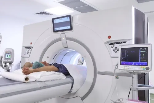
Medical imaging methods play an important role in the diagnosis and treatment planning of many diseases. At Yeditepe University Hospitals Imaging Unit, diagnostic imaging services are provided using state-of-the-art imaging technologies and a team of specialized physicians.
What is the Imaging Unit?
The Imaging Unit is a department that contributes to the evaluation and diagnosis of diseases using various imaging methods.
Our unit is equipped with advanced technological devices, including:
- 3 Tesla Magnetic Resonance Imaging (MRI),
- Multi-Slice Computed Tomography (MD-CT),
- PET/CT,
- Gamma Camera-SPECT,
- Flat Panel Digital Angiography (DSA),
- Digital Fluoroscopy,
- Digital Direct Radiography (X-ray),
- Digital Mammography,
- Bone Densitometry,
- 4-D Ultrasonography,
- Doppler Ultrasonography,
- Portable X-ray,
- C-Arm.
Digital Imaging and Archiving Infrastructure
All images are acquired digitally, collected in a central system, stored, evaluated, and archived with their reports in digital format. They are then transmitted via the intranet network to all units of our hospital. Through this computer and internet network, our Imaging Unit can also connect with other imaging centers around the world and exchange information.
The PACS (Picture Archiving & Communication System) and RIS (Radiology Information System) required for this function are used in our hospital with extensive capabilities.
Diagnostic and Interventional Radiology Procedures
Our goal is to provide accurate diagnoses in a short time, guided by current knowledge, with our experienced radiology and nuclear medicine specialists, to communicate these results rapidly to the relevant physicians, and to contribute to our patients' treatment processes.
In addition to diagnosis, minimally invasive treatment methods are also performed in our Interventional Radiology Unit using state-of-the-art technologies.
Molecular Imaging Technologies
There have been revolutionary changes in the field of imaging. Evaluating images alone or attempting to diagnose based solely on the morphological appearance of organs and tissues is no longer sufficient. Our clinicians expect definitive diagnoses based on quantitative data from imaging units. Today, with molecular imaging techniques, in addition to morphological images of organs, quantitative information can be obtained at the cellular and even molecular level regarding the metabolic and functional characteristics of pathological tissues and tumors.
With PET/CT in our unit, the increased metabolic activity of tumors is evaluated and their localization is determined. Functional MRI, along with MR Proton and Phosphorus Spectroscopy, allows the identification of hypervascularized areas within tumors during imaging. The types and quantities of molecules within tumors can be assessed, enabling sub-classification and grading of tumors. In light of this information, the diagnosis of diseases can be made more reliably. Furthermore, the effectiveness of applied treatment can be monitored at the molecular level by evaluating the metabolic activation and chemical and physical changes of the tumor. The need for biopsy for diagnosis is gradually decreasing. It is predicted that this invasive and time-consuming method may eventually be completely eliminated, in line with advances in genetics, molecular biology, and imaging technologies. Functional MRI also enables imaging of vital functional areas in the brain, allowing for maximum surgical resection of brain tumors without damaging these critical regions.
Research and Scientific Studies
As befits a university hospital, a true research environment is being established, and contributions are being made to the development of the Molecular Imaging Research Unit, whose project is in its final stages and which has been established for the first time in Turkey at Yeditepe University Hospital.
Objective of the Imaging Unit
Our goal as Yeditepe University Hospitals Imaging Unit is to apply current imaging techniques, develop new technologies, and offer them for the benefit of our country and all humanity, competing with leading universities worldwide.
General Preparation Forms
- Patient Information Form
- MRI Preparation Form
- Pregnancy Information and Consent Form for Radiological Examinations
This content was prepared by Yeditepe University Hospitals Medical Editorial Board.
”
See Also
- Anterograde Pyelography, Nephro Cystography, Anterograde Urethrography
- Stomach-Duodenum X-ray
- Intravenous Pyelography (IVP)
- Enteraclysis (Small Intestine)
- Imaging Center / Education and Research
- Patient Preparation Information
- Dacryocystography (Tear Sac and Tracts)
- Pediatric CT
- Pediatric MRI
- Neuro Radiology
- Magnetic Resonance Imaging (MRI)
- Multi Slice CT
- Soft Tissue Ultrasonography
- Extremity CT
- Ultrasound and Doppler Ultrasonography
- Fluoroscopy
- Digital X-ray
- Cystography, Retrograde Cystography, Fractionated Cystography (Progressive Cystography)
- Voiding Cystography, Voiding Cystourethrography, (Micturition Cystourethrography)
- TRANSVAGINAL US and Doppler Us
- Cyanography (Salivary Glands)
- Portal and Main Vascular Doppler Us
- Colostomy, Ileostomy Radiography
- Colon X-ray (Single or Dual Contrast)
- Hysterosalpingography (HSG)
- T-Tube Cholangiography (Biliary Tract)
- Lower Abdomen CT
- Radiology of Abdomen
- Oral Cholecystography (Gallbladder and Pathways)
- Renal Doppler Us
- Virtual Colonoscopy CT
- TR Doppler Us - TRUS
- Whole Abdomen CT
- Upper Abdomen CT
- Coronary CT Angiography (Virtual Angiography)
Alo Yeditepe
Our Branches With This Unit