Alo Yeditepe

Interventional Radiology
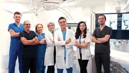
The effective use of imaging methods in diagnosis and treatment processes forms the basis of interventional radiology. At Yeditepe University Hospitals Department of Interventional Radiology, diagnostic and therapeutic procedures for many diseases are performed through image-guided interventional procedures.
What Is Interventional Radiology?
Interventional Radiology is one of the subspecialty areas of radiology that utilizes medical imaging methods in the diagnosis and treatment of diseases. Interventional radiologists not only make diagnoses using imaging techniques but also play an active role in the treatment process of diseases.
What Are Interventional Radiology Procedures?
Interventional radiology procedures are carried out in different areas, including diagnostic, vascular, and therapeutic interventions. In this context, numerous interventional procedures for diagnosis and treatment are performed under image guidance.
I. Diagnostic Applications
Biopsies
Fine-needle aspiration biopsies or Tru-cut biopsies are performed under the guidance of various radiological imaging techniques (Ultrasonography and/or Computed Tomography) for:
- Neck masses and Thyroid nodules
- Thoracic masses
- Intra-abdominal organ biopsies (Liver, pancreas, kidney)
- Bone biopsies
Arteriography Applications
This is a method of imaging blood vessels (arteries and veins) using X-rays with the administration of contrast agents. Usually, access is gained through the femoral artery and, rarely, the axillary artery, using a needle and catheter to deliver contrast material to the desired region.
Angiographic Applications
- Carotid - Cerebral (neck/brain) angiography
- Aortic arch angiography, Thoracic (chest) angiography
- Abdominal aortography
- Renal (kidney) angiography
- Celiac / SMA (abdominal organs) angiography
- Upper/lower extremity arteriography
Angiography (Arteriography) Applications
Thanks to high-technology angiography devices, high-resolution image quality allows the most distal branches to be displayed with great clarity, and 3D imaging can also be obtained.
Abdominal Aortography, Extremity Angiographies
The arterial circulation of the distal abdominal aorta, legs, and feet is visualized.
Renal Angiography
Renal artery stenosis, which can be a cause of hypertension, is investigated using this method. It is also performed in patients with suspected kidney tumors, as well as before and after renal transplantation to examine vascular structures.
Celiac/SMA (Superior Mesenteric Artery) Angiography
Vascular pathologies of the stomach, duodenum, liver, spleen, and pancreas can be diagnosed. If a gastrointestinal bleeding source is being investigated, treatment is usually performed together with angiography.
Thoracic Angiography
The thoracic aorta is examined for dissection and aneurysm.
Aortic Arch Angiography
Stenosis, dilatation, and occlusions of the aortic arch and brachiocephalic vessels are diagnosed.
Upper Extremity Angiography
The vascular structures of the arms are evaluated. If obstructions or stenoses are detected, interventional radiological methods are applied for treatment.
Carotid/Cerebral Angiography
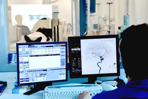
In these examinations, stenoses, occlusions, aneurysms, and tumors of the vascular structures of the head and neck region are diagnosed. 3D images of aneurysms can also be created, and the relationship between the aneurysm, parent artery, and neck can be accurately determined before surgery or embolization.
- Cerebral Angiography
- 3D Cerebral Angiography
- Pulmonary Angiography: Performed to investigate pulmonary hypertension and embolism. The access site in this method is the femoral vein.
Venography Applications
- Superior and inferior venacavography
- Pulmonary angiography
- Renal venogram, Adrenal gland venography
- Upper/lower extremity venography
Inferior Venacavography: Structures of the inferior vena cava, tumor blockages, and causes of thrombosis are revealed.
Renal Venogram: Thrombosis in the renal veins is investigated.
Adrenal Venography: Performed to visualize adrenal gland tumors.
Leg Venography: Deep veins of the leg and abdomen are investigated for thrombosis.
Saphenous Vein Venography: Performed to evaluate the saphenous vein to be used as a graft before bypass surgery.
II. Therapeutic Applications
Thoracic Interventions: Nonvascular Applications
- Thoracentesis
- Abscess drainage
- Stent applications for tracheobronchial stenosis
Abdominal Interventions
- Biliary tract interventions (percutaneous transhepatic cholangiography [PTC], biliary drainage, biliary stenting)
- Biliary stent placement
- Drainage of renal cysts and abscess collections
- RF Ablations
- Percutaneous nephrostomy
- Abscess and collection drainage
- Antegrade ureteral stenting
- Biliary Stenting
- Renal Interventions
III. Vascular Applications
Arterial Applications
- Treatment of vascular stenoses throughout the body with balloons and stents
- Carotid Angioplasty and Stenting
- Cranial stenting
- Subclavian and vertebral artery stenting
- Renal Artery Angioplasty and Stenting
- Mesenteric Artery Angioplasty
- Iliac, Femoral, and Popliteal artery angioplasty
- Embolization of cranial aneurysms and arteriovenous malformations (AVMs)
- Intra-arterial thrombolysis in stroke cases
- Carotico-cavernous fistula embolization
- Embolization for epistaxis, hemoptysis, and acute gastrointestinal bleeding
- Aortic aneurysm endograft applications
- Cranial and Neck Tumor Embolization
Treatment of Carotid and Intracranial Stenoses with Balloon Angioplasty and Stenting
Intra-arterial Thrombolysis in Acute Stroke
In acute stroke cases, the cause may be thrombotic occlusions of the major cerebral arteries. Such pathologies can be treated within a specific time limit.
Embolization of Cerebral Aneurysms
Anterior and posterior circulation aneurysms can be embolized with direct coiling or balloon-assisted coiling.
Embolization of Cranial and Extracranial Arteriovenous Malformations and Fistulas
AVM and AVF pathologies can be embolized using various embolic agents.
Balloon Angioplasty and Stenting of Brachiocephalic, Subclavian, Aortoiliac, Femoral, and Popliteal Stenoses and Occlusions
Peripheral artery stenoses and occlusions can be treated with balloon and stent applications to restore arterial patency and relieve patient symptoms.
Endograft Treatment of Aneurysms and Pseudoaneurysms in Peripheral Vascular Structures
With the application of covered stents, aneurysms in the thoracic and abdominal aorta, as well as pseudoaneurysms developing in the femoral and other arteries, can also be treated.
Balloon and Stent Applications in the Treatment of Renal Stenoses
In the treatment of hypertension, renal artery stenoses can be treated with stents and balloons to restore patency.
Liver and Kidney Chemoembolization and Uterine Fibroid Embolization
In the treatment of hepatocellular carcinoma, intra-arterial chemoembolization can prolong patient survival. Embolization is an important alternative to surgery in the treatment of uterine fibroids.
Venous Applications:
- Angioplasty and stenting of venous stenoses
- Hemodialysis catheter placement
- Angioplasty for hemodialysis fistula stenoses
- Inferior Vena Cava filter placement
- TIPS (Transjugular Intrahepatic Portosystemic Shunt)
- Uterine fibroid embolization
- Venous catheter placement
Other Interventional Radiological Treatments
Diabetic Foot Treatment
Stenoses in the foot vessels of diabetic patients can be treated with balloon and laser angioplasty, with good results.
Oncological Applications and Treatments
- Venous port placement
- Permanent tunneled and temporary venous catheter placement
- Liver tumors:
- RF ablation
- Transarterial chemoembolization
- Portal vein interventions
Treatment of Carotid and Intracranial Stenoses with Balloon Angioplasty and Stenting
Intraarterial Thrombolysis in Acute Stroke
In acute stroke cases, the cause may be thrombotic occlusions of the main cerebral arteries. Such pathologies can be treated within a certain time limit.
Cerebral Aneurysm Embolization
Anterior and posterior circulation aneurysms can be embolized by direct coil or balloon modeling.
Cranial and Extracranial Arteriovenous Malformation and Embolization of Fistulas
AVM and AVF pathologies can be embolized using various embolizing agents.
Balloon Angioplasty and Stenting of Brachiocephalic, Subclavian, Aortoiliac, Femoral and Popliteal Stenoses and Occlusions
Peripheral artery stenosis and occlusions can be eliminated by ensuring the patency of the arteries with balloon and stent applications.
Endograft Treatment of Aneurysms and Pseudoaneurysms in Peripheral Vascular Structures
With the application of covered stents, aneurysms in the thoracic and abdominal aorta, and pseudo-aneurysms in the femoral and other arteries can also be treated.
Balloon and Stenting in the Treatment of Renal Stenosis
In the treatment of hypertension, renal artery stenosis can be treated with a stent and balloon, which can be cleared.
Liver, Kidney, Chemoembolization, and Uterine Myoma Embolization
In the treatment of hepatocellular carcinomas, the survival of the patient can be prolonged with intraarterial chemoembolization. Embolization is an important alternative to surgery in the treatment of uterine myomas.
Venous applications:
- Angioplasty and stenting of venous stenosis
- Hemodialysis catheter placement
- Treatment of hemodialysis fistula stenosis with angioplasty
- Placing a filter in the Vena Cava Inferior
- TIPS
- Uterine fibroid embolization
- Venous catheter placement
Other Interventional Radiological Therapies
Diabetic Foot Treatment
Stenosis in the foot veins of diabetic patients can be treated with balloon and laser angioplasty and good results are obtained.
Oncological Applications and Treatments
Venous Port Placement
Permanent tunneled and transient venous catheter placement
In liver tumors;
- RF ablation
- Trans arterial chemoembolization
- Portal vein interventions
This content was prepared by Yeditepe University Hospitals Medical Editorial Board.
”
See Also
- Radioembolization in Liver Tumor Treatment
- Diagnosis and Treatment of Acute Stroke
- The Cause of Abdominal Pain Has Not Been Found for Years... Varicose Veins Appeared in Her Ovary
- Interventional Methods in the Treatment of Acute Stroke
- Breast MRI
- Interventional Radiology is Used in Many Fields
- What Diseases Is Interventional Radiology Used For?
- What is a Brain Aneurysm?
- Interventional Oncology
Alo Yeditepe