Alo Yeditepe

What Is Astigmatism? What Causes It? What Are Its Symptoms and Treatment?
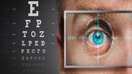
Astigmatism is a common vision defect that arises because the eye cannot focus light correctly. When the curvature of the cornea, the transparent layer at the front of the eye, or of the lens of the eye differs from normal, light cannot be focused properly on the retina. As a result, both near and distant objects may appear blurred or distorted.
Astigmatism, which can occur at any age, may lead to eye strain, headaches and vision problems, especially when driving at night, if left uncorrected. Astigmatism symptoms, which can significantly affect daily life and quality of vision, can be brought under control with glasses, contact lenses or, in suitable cases, surgical methods, depending on the person's eye structure and needs. You can find questions frequently asked by the public, such as "What Does Astigmatism Mean?", "How Does Astigmatism Go Away?", and much more in our article below.
What Is Astigmatism?
Astigmatism is a refractive error that arises because the curvature of the cornea or the lens of the eye is not the same in all directions. The cornea, which normally has a nearly round shape, may in astigmatism have a steeper curvature in one direction and a flatter curvature in the other. The difference in curvature of the cornea or lens prevents light from being refracted evenly within the eye, causing the image to be perceived as blurred or distorted. People with astigmatism may have difficulty seeing both near and distant objects clearly.
For vision to take place, light reflected from objects must pass through the structures of the eye and be focused on the retina. Light particles coming into the eye from outside first enter the eye by being refracted at the cornea, the transparent layer resembling a watch glass located at the very front of the eye. The refracted light passes through the clear fluid behind the cornea and the pupil to reach the lens of the eye. Refracted again here, the light passes through the vitreous body, the clear, gel-like substance that fills the inside of the eye, and reaches the retina at the back of the eye. Light-sensitive cells in the retina convert the light into nerve signals and transmit them to the brain via the optic nerve. The brain processes this information and enables us to see.
The term astigmatism derives from the Greek words “a”, meaning absence, and “stigma”, meaning point. In normal vision, the cornea and the lens of the eye work together so that light forms a sharp image on the retina. In astigmatism, however, because the curvature of one or both of the cornea and the lens varies in different directions, light cannot be refracted evenly. Since light cannot be focused sharply at a single point, the image may be perceived as blurred or distorted.
What Are the Types of Astigmatism?
Astigmatism is a refractive error that develops as a result of the cornea or the lens of the eye being more steeply curved in one direction. According to which structure of the eye the refractive error originates from, astigmatism is divided into two classes: corneal astigmatism and lenticular astigmatism. In corneal astigmatism, the cornea has an oval shape rather than its normal round shape. In lenticular astigmatism, there is a shape abnormality in the lens of the eye.
In some cases, astigmatism may also be classified as regular (normal) astigmatism and irregular astigmatism. In regular astigmatism, the curvature of the cornea in one direction is different from that in the other direction. However, because this difference follows a certain pattern, how light is refracted is relatively predictable. For this reason, regular astigmatism can usually be corrected with astigmatism lenses or glasses.
In irregular astigmatism, the shape of the cornea is more complex; that is, the curvatures in different regions of the surface are not consistent with one another. This causes light to be refracted in a more complex way within the eye. As a result, compared with regular astigmatism, it may be more difficult to fully correct the blurring of the image with standard glasses. Irregular astigmatism may arise from diseases affecting the structure of the cornea, such as keratoconus, or from factors such as eye trauma affecting the cornea.
What Are the Symptoms of Astigmatism?
When astigmatism symptoms are mentioned, the first thing that comes to mind is objects at all distances appearing blurred. Vision is blurred because, due to refractive errors of the eye, the image cannot be focused precisely on the retina. Here, it is important where the vision defect originates from. Refractive errors are generally examined in four groups: presbyopia, hyperopia, myopia and astigmatism.
In myopia, a refractive error that develops as a result of the eye being longer than normal from front to back, people cannot see distant objects clearly. In hyperopia, which results from the eye being shorter than normal, near objects appear blurred. Astigmatism, unlike these, causes objects at all distances to appear blurred.
Under normal conditions, the cornea and lens at the front of the eye have an equally round shape. This allows light particles to be focused sharply on the retina. In astigmatism, however, the cornea or lens having a relatively oval shape causes light to fall in front of or behind the retina. This makes it difficult to see both distant and near objects. Accordingly, the main astigmatism symptoms can be listed as follows:
- Seeing objects at all distances as blurred, distorted and hazy,
- Impaired depth perception,
- A constant need to squint in order to see better,
- Headaches that cannot be explained by another cause,
- Focusing problems,
- Eye strain,
- Seeing halos or glare when looking at a light source, especially at night,
- General fatigue.
Over time, astigmatism may also lead to the development of amblyopia (lazy eye), especially in children. For this reason, it is recommended that children have an eye examination at least once between the ages of 3 and 5, and once a year after the age of 6.
Although the listed symptoms are commonly encountered in astigmatism, since conditions such as headache and impaired depth perception may also result from other health problems, it is important first to consult a specialist doctor and have the necessary check-ups done.
How Does a Person with Astigmatism See?
People with astigmatism may see objects as blurred, hazy, distorted or shadowed. For example, when reading a book, the letters may run into each other, faint shadows may form at the edges of the letters, or the text may appear to overlap. Straight lines may be perceived as sharper in some directions and blurred or elongated in others. When driving at night, the headlights of oncoming vehicles and street lamps may appear brighter than normal, elongated into streaks, or surrounded by rings of light.
How Does Astigmatism Affect Night Vision?
Astigmatism may also cause impairment, particularly of night vision. For example, the light from car headlights may glare excessively, and halos or streaks may be seen around street lamps at night.
In fact, the eye's normal working mechanism underlies this. When a large amount of light enters the eye, the iris muscles contract and constrict the pupil to reduce the amount of light entering. In the dark, on the other hand, the pupil dilates in order to make better use of the small amount of light in the surroundings. In this way, more light enters the eye and reaches the retina.
In people with astigmatism, this causes light to pass through a wider and asymmetric part of the cornea outside its centre. As a result, especially when driving at night, the headlights of oncoming vehicles and the light of street lamps may appear scattered, elongated into streaks or surrounded by rings of light (halos). In addition, shadowed or double images may occur at the edges of objects. The reduction in image sharpness and contrast perception can make it difficult to distinguish distant objects, lanes and details on the road, adversely affecting the driver's estimation of distance (depth perception).
What Causes Astigmatism?
Although the causes of astigmatism are not definitively known, genetic factors are thought to play an important role. The most common cause of astigmatism is that the curvature of the cornea, located at the very front of the eye, is not the same in different directions. The cornea, which normally has a nearly round shape, may be steeper in one direction and flatter in the other. The difference in the shape of the cornea prevents light from being refracted evenly within the eye and impairs the sharpness of the image. In rarer cases, differences in the shape of the lens of the eye may also lead to astigmatism.
Family history plays an important role in the emergence of astigmatism. Some studies on astigmatism in preschool children show that astigmatism is more common in children whose mother and/or father has astigmatism. However, astigmatism is not only congenital.
Factors such as certain health problems that can distort the shape of the cornea or eye trauma may also cause astigmatism to develop later in life. For example, keratoconus, characterised by the cornea losing its normal round shape and bulging outward into a cone shape, can disrupt the smooth shape of the cornea and lead particularly to irregular astigmatism. Corneal injuries or certain infectious diseases that cause damage to the cornea may also change the way light is refracted, that is, cause astigmatism. The main causes of astigmatism and risk factors are as follows:
- Genetic predisposition,
- Eye trauma,
- High myopia or hyperopia,
- Conditions such as keratoconus or lenticonus,
- Complications of eye surgery, such as cataract surgery.
The corneal layer has a flexible structure. In addition to the above, structurally excessive pressure of the eyelid on the cornea or eyelid diseases such as chalazion (eyelid cyst) may lead to a change in the shape of the cornea and therefore to astigmatism.
Why Does Astigmatism Increase?
The degree of astigmatism may increase or decrease over time due to age-related changes in the structure of the eye. Changes in the curvature of the cornea and lens, eye trauma and health problems affecting the structure of the cornea, such as keratoconus, may also cause changes in the degree of astigmatism.
Does Astigmatism Cause Headaches?
Astigmatism may cause headaches, particularly after prolonged reading, computer use or tasks requiring concentration. The eye's inability to focus light sharply may lead the person to squint in order to see the image more clearly and to strain while focusing. As a result, pain around the eyes, eye strain and headaches may occur.
Should People with Astigmatism Wear Glasses All the Time?
Not everyone with astigmatism needs to wear glasses all the time. Whether glasses are to be worn constantly or only for certain activities is determined according to the degree of astigmatism, visual complaints and the person's age.
How Is Astigmatism Diagnosed?
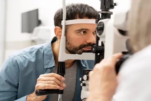
A diagnosis of astigmatism can only be made by measurements performed during a comprehensive eye examination. During the examination, specialists first listen to the person's complaints and take a family history. They then examine visual acuity and the way light is focused in the eye with the help of various devices and tests. In this way, it can be determined whether blurred vision is due to astigmatism or another refractive error. The main methods used in the diagnosis of astigmatism are the visual acuity test, the refraction test and keratometry.
Visual Acuity Test
One of the methods commonly used during an eye examination is the visual acuity test. One of the methods used to measure visual acuity is the letter chart, also known as an eye chart or Snellen chart. During the examination, the person is asked to look at the chart from a distance of approximately 6 metres and read letters of different sizes. At this stage, each eye is assessed separately. First one eye is covered and the person is asked to read the letters with the uncovered eye, then the other eye is tested. The letters get smaller towards the lower lines, and the line down to which the person can read is assessed.
In people who are illiterate or who cannot complete the letter test for any reason, the E chart can be used. In this method, the person is asked which way the arms of the letter E, facing in different directions, are pointing. The person answers by stating the direction of the letter or by showing it with their hand. In young children, charts of shapes or symbols suitable for their age and communication skills can also be used in the diagnosis of astigmatism.
Visual acuity gives an idea of whether the person has a vision problem. However, this test may not be sufficient for determining the degree of astigmatism or the type of refractive error in the cornea or lens. To confirm the diagnosis of astigmatism, additional investigations such as a refraction test may be needed in addition to the visual acuity test.
Refraction Test
The refraction test is an eye examination performed to determine, by measuring how the eye refracts light, whether the person needs to use glasses or contact lenses and the lens prescription they need. This test, used to detect refractive errors such as astigmatism, myopia and hyperopia, is an important part of a routine eye examination.
During the test, how light is refracted as it passes through the cornea and lens is examined first. This measurement is usually performed with the help of a computerised device. In computerised measurement, the device analyses the light reflected from the retina to examine whether there is a refractive error in the eye. 20/20 visual acuity refers to normal distance visual acuity under standard test conditions; however, it does not by itself show that there is no refractive error. If the test result is not 20/20, this may mean that there is a refractive error such as myopia, hyperopia or astigmatism.
Measurements are then taken with different lenses to determine the glasses or contact lens prescription the person needs. While looking at the letter chart in front of them, the person covers one eye and tries to read the smallest letters they can see with the other eye. The ophthalmologist, using different lenses, asks with which option the letters are seen more clearly. The same procedure is repeated for the other eye. Based on the results obtained, the lens prescription that will best correct the person's vision is determined.
In addition to astigmatism, the refraction test also helps identify other refractive errors such as myopia and hyperopia. In addition, data that enable the detection of certain eye diseases that may cause vision problems can also be obtained.
Keratometry Test
The keratometry test is an eye examination performed to measure the curvature of the cornea. Since astigmatism can arise from the cornea having a curvature different from normal, the test helps in identifying astigmatism.
During the test, a device called a keratometer is used. The person places their chin on the device's rest, leans their forehead against the relevant part and looks at the images inside the device. The ophthalmologist aligns the images and takes curvature measurements in different regions of the cornea. The same procedure is repeated for the other eye. No special preparation is needed for this short and painless procedure.
In keratometry measurement, the curvature values in the steepest and flattest regions of the cornea are determined. While corneal curvature is more regular in eyes without astigmatism, a difference between these values may be seen in people with astigmatism. The measurements obtained help the ophthalmologist understand the cause of the vision defect and plan the appropriate treatment.
The keratometry test can also be used to determine contact lens fit and to calculate the power of the intraocular lens before cataract surgery. However, since it measures the curvature of the cornea approximately, it does not show every detail on its own. When necessary, more detailed imaging methods such as corneal topography or corneal tomography can also be used.
Is Astigmatism Dangerous?
If left untreated, astigmatism may cause various complications. For example, if there is astigmatism in one of the eyes or the astigmatism in one eye is more severe than in the other, problems such as amblyopia (lazy eye) may develop if it is not treated.
What Is the Treatment for Astigmatism?
Astigmatism treatment is planned according to the degree of the refractive error in the eye, the problems the person experiences in daily life and their preferences. Treatment may not be necessary in people with mild astigmatism who do not have vision problems. However, in astigmatism that causes complaints such as blurred vision, glasses, contact lenses or, in suitable individuals, surgical methods may be used.
Glasses and Contact Lenses
Glasses and contact lenses are the most commonly used methods in the treatment of astigmatism and do not require a surgical procedure. Glasses or contact lenses prepared according to the person's eye measurements can reduce blurred vision and increase visual acuity by enabling light to be focused more accurately on the retina.
Cylindrical spectacle lenses used to correct astigmatism help light to be focused correctly on the retina. Contact lenses are also devices designed to have a similar effect to glasses. The main difference is the way they are used. For people who prefer contact lenses, coloured lens options suitable for correcting astigmatism are also available. However, when choosing coloured lenses for astigmatism, it is important to choose products appropriate to the eye structure and prescription. Which method will be used is determined by the ophthalmologist according to the degree of astigmatism, the person's eye structure and daily needs.
Ortho-K (Orthokeratology) Treatment
Orthokeratology is a method in which special rigid contact lenses are used to temporarily change the shape of the cornea. These lenses can be worn during sleep and removed in the morning. Thanks to the temporary change in corneal curvature, the person can see more clearly during the day without using glasses or contact lenses.
However, the vision correction provided by orthokeratology is not permanent. When lens use is discontinued, the cornea gradually returns to its previous shape and the vision defect reappears. For this reason, for the effect of the treatment to continue, the lenses must be used in accordance with the physician's recommendation.
Laser Treatment of Astigmatism
Laser astigmatism surgery can be performed in suitable individuals to correct astigmatism permanently. In these procedures, the curvature of the cornea is reshaped with the help of a laser, with the aim of focusing light more accurately on the retina. However, not every astigmatism patient is suitable for laser treatment. Before a decision is made, the corneal structure, the degree of astigmatism and general eye health are examined in detail.
The main surgical methods used in the treatment of astigmatism are as follows:
- LASIK: A thin flap is created on the cornea. The shape of the cornea is then corrected with a laser and the flap is repositioned.
- PRK: After the epithelial layer on the corneal surface is removed with the help of a laser, special instruments or a special solution, the cornea is reshaped with a laser.
- LASEK: The epithelial layer on the corneal surface is loosened and moved aside, and the underlying tissue is reshaped with a laser. After the procedure, the epithelial layer is repositioned.
- SMILE: A small, lens-shaped piece of tissue (lenticule) is created with a laser within the corneal tissue and removed through a small incision. In this way, the shape of the cornea is changed.
Astigmatism laser treatment prices and similar details are clarified before the operation. In addition, as with any surgical procedure, there may be risks of infection, changes in quality of vision and other complications after laser treatment. For this reason, before the treatment is chosen, the specialist ophthalmologist also informs patients about the possible risks.
Astigmatism is a common refractive error that can lead to blurred vision, usually due to differences in curvature of the cornea and rarely of the lens of the eye. Quality of vision can be improved with appropriate glasses, contact lenses or, when necessary, surgical methods. To determine the degree of astigmatism and choose the treatment suited to the eye structure, it is important not to neglect regular eye examinations. Specialists recommend that, even without any complaints, healthy adults under the age of 60 have an eye examination every 2 years, and those over 60, particularly those with a family history of a health problem such as glaucoma, once a year.
If you notice symptoms such as blurred vision or loss of sharpness, or if you would like to have a routine eye examination, you can consult the specialist physicians at the Ophthalmology department of Yeditepe University Hospitals to have the necessary check-ups and obtain information about the treatment options suitable for you.
Frequently Asked Questions About Astigmatism
What Happens When You Have Astigmatism?
When you have astigmatism, problems such as blurred vision, eye strain, headaches and lights appearing scattered or elongated, especially at night, may occur.
What Is Astigmatism in Babies?
Astigmatism in babies is a vision defect that arises because light cannot be focused sharply on the retina due to differences in the shape of the cornea or the lens of the eye. Since babies' visual system continues to develop after birth, their visual abilities change over time in the first months. However, early detection of refractive errors such as astigmatism is important for healthy visual development.
Can Astigmatism Be Corrected with Natural Treatment Methods?
There is no natural treatment method scientifically proven to eliminate astigmatism. In other words, astigmatism cannot be corrected with natural treatment methods. Eye exercises, herbal products or dietary changes cannot correct the curvature of the cornea or the lens of the eye. To correct the vision defect, glasses, contact lenses or, in suitable individuals, surgical methods can be used.
What Glasses Prescription Is Needed for Astigmatism?
The glasses prescription required for astigmatism is determined after an eye examination. The astigmatism-related part of the prescription consists of the cylinder (CYL), which shows the degree and extent of the irregularity, and the axis (AX), which shows the angle at which the astigmatism lies.
Can an Astigmatic Eye Be Corrected?
An astigmatic eye can be corrected with surgery in suitable patients. Glasses or contact lenses cannot completely correct the problem, but they can provide clearer vision.
Which Lens Corrects Astigmatism?
Although astigmatism cannot be completely corrected with glasses, cylindrical spectacle lenses or toric contact lenses are generally used to achieve clearer vision.

About
Faculty and Year of Graduation:
Istanbul University Cerrahpaşa Faculty of Medicine, 1981
Alo Yeditepe