Alo Yeditepe
Alo Yeditepe
Eyelid Diseases
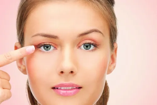
It is a condition where the upper eyelid is lower than it should be. Although the interval between the upper and lower eyelids varies between individuals, it is about 10 -11 mm on average and the upper eyelid covers the colored part of the eye by 2 mm.
Operating under Yeditepe University Hospitals, the Eye Center is an eye diseases center that trains students and assistants in the Department of Ophthalmology and provides services in 15 different branches.
Eyelid Drooping
It is the condition that the upper eyelid is located lower than it should be. The gap between the upper and lower eyelids varies between individuals, but it is about 10-11 mm on average and the upper eyelid covers the colored part of the eye by 2 mm. When it covers more than this or the difference between the two eyes reaches a remarkable size, droopy eyelids can be taken into consideration. This condition can be unilateral or bilateral. While mild drooping creates only an aesthetic defect, more severe ones can also affect vision.
What is the Cause of Eyelid Drooping?
Eyelid drooping (ptosis) can be congenital, or it can be caused by injury, muscle, and nerve diseases, especially with the relaxation of the muscle that lifts the eyelid due to age.
Various eye surgeries and the use of contact lenses can also cause drooping of the eyelid due to the relaxation of this muscle.
How Is Eyelid Drooping Treated?
The treatment of eyelid drooping is surgery. In the procedure, it is aimed to lift the eyelid by strengthening the levator muscle. Eyelid drooping surgery is performed according to the type and weight of the droopy eyelid. There are different techniques. It can be performed from the skin fold or under the upper eyelid with scarless surgery. In congenital ptosis, if the functioning of the levator muscle that lifts the upper eyelid is low, the muscle in the eyebrow area of the eyelid is suspended with the help of a silicone strip (frontale suspension method). Your doctor determines the method to be applied in surgery according to the examination findings of the eyelid. Ptosis surgeries performed in adults are generally performed under local anesthesia. The patient is discharged on the same day and applying ice on the day of surgery prevents the development of swelling and bruising.
Lower Eyelid Entropion
Inward turning of the eyelid margin may be congenital or may develop later. It is mostly related to aging. Causes also include microbial diseases such as trachoma, chronic conjunctivitis, and chemical and physical injuries. Contact of the eyelashes with the surface of the eye causes complaints such as redness, tearing, stinging, and inability to look at the light. In advanced cases, wounds or even punctures can be seen on the surface of the eye. The treatment is surgery. Entropion surgery can also be performed congenitally in newborns. However, it requires surgical experience.
Lower Eyelid Ectropion
The outward turning of the eyelid margin also often occurs as a result of aging. It can also develop congenitally, after paralysis of the nerve closing the eyelid (facial paralysis) and injuries. Eye-opening, redness, burning, and watering are the most common symptoms. This problem can also be treated surgically.
Chalazion
As a result of the occlusion of the sebaceous glands opening to the edge of the eyelid, the contents accumulate on the lid. First, acute inflammation similar to a stye occurs, followed by cystic formation. This condition, called a chalazion, is painless. Extremely large ones can cause astigmatism and affect vision. Rarely, they can be confused with eyelid cancers. As in the treatment of styes, hot compresses on the eyelid, antibiotics, and cortisone eye drops or pomades are used. Surgical intervention is used in cysts that do not disappear with this treatment.
This content was prepared by Yeditepe University Hospitals Medical Editorial Board.
”
See Also
- What is Cataract? Symptoms and Treatment
- Do not Risk Going Blind While Trying to Have Colored Eyes
- Watch Out Blue Light Emitted By Devices For The Health of Your Eyes
- Pollen is Now Seen Outside of Seasonal Changes
- Pay Attention to the Flashing Light in the Eye!
- Medication Administration to the Eye
- Thyroid-Related Eye Diseases
- Glaucoma
- If You Have Diabetes, Consult An Ophthalmologist At Least Once A Year.
- It was Told that She could Lose Both Eyes, but She Recovered with an Operation
- Untreated Strabismus Can Cause Vision Problems!
- The Danger to the World: MYOPIA
- Is Strabismus Genetic?
- One in 20 Newborns Have Tear Duct Obstruction
- Are Flies Flying Before Your Eyes?
- 7 Tips to Protect Your Eye Health
- Allergic Conjunctivitis (Eye Allergy)
Alo Yeditepe


