Alo Yeditepe
Alo Yeditepe
Imaging Methods During Pregnancy
The Most Beautiful Journey
Pregnancy is the most incredible journey in a woman's life. In this journey that lasts for 9 months, both your body and soul pass through various stages and proceed toward the last station. The person who will greet you at the end of this road is the "child", the equivalent of indescribable love on earth.
With ultrasonographic examinations performed at certain weeks of pregnancy, important data can be obtained for both the baby's and the mother's health.
Prof. Dr. Rukset Attar, Yeditepe University Kozyatağı Hospital Gynecology, Obstetrics, and IVF Specialist, made important statements by emphasizing that pregnancy can be examined in great detail thanks to ultrasonography devices.
What is the Place and Importance of Ultrasonography During Pregnancy?
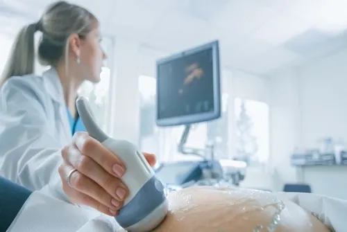
Ultrasonography plays an important role in the diagnosis and follow-up of pregnancy. We can determine whether the pregnancy is an "intrauterine pregnancy" or an "ectopic pregnancy" by ultrasonographic examination. We can also detect whether the pregnancy is singleton or multiple pregnancies by ultrasonography. It also gives information about the development of the baby, anomalies, the amount of water in which the baby develops, which we call "amniotic fluid", and the location and condition of the partner of the baby, which we call "placenta".
Is Ultrasound Harmful to the Baby?
There is no scientific evidence that ultrasound harms the baby. However, the effects of heat increase during an ultrasound are not clear. As a result of scientific studies, it has been reported that a temperature increase of 1.5 degrees is not harmful to the baby.
How Does Ultrasound Work?
Ultrasound works with sound waves. Sound waves are sent to the tissue with the help of a device (ultrasound probe). Sound waves coming into the tissue are reflected and the reflected sound waves are displayed on a screen.
How Often Should Ultrasound Be Performed?
Ultrasound is performed in the first diagnosis of pregnancy for nuchal translucency and anomaly scanning at 11-14 weeks, for anomaly scanning between 18-22 weeks, in the last month of pregnancy, and in other periods deemed necessary by your physician.
What Is Detailed Ultrasound and When Is It Performed?
Detailed ultrasound is performed between 18-22 weeks by a perinatology specialist specialized in risky pregnancies, congenital disabilities, and diseases.
Should It Be Done in Multiple Pregnancies?
Ultrasonography must be performed in the first trimester to determine whether the pregnancy is multiple or not.
What Does the Report Contain?
The ultrasound report includes information about the mother's age, last menstrual period, expected date of birth, placenta location, amount of amniotic fluid, baby's heartbeat, number of vessels in the baby's umbilical cord, information about the baby's development, blood flow, and cervical length. In addition, suggestions are also given when necessary.
Can Every Anomaly Be Detected with Detailed Ultrasound? Is Detailed Ultrasound Alone Sufficient for the Diagnosis of Disabilities?
Not every anomaly can be detected with detailed ultrasound. With the detailed ultrasonography examination, it is accepted that the risk of Trisomy 21 is reduced by 60-70 percent, and the risk of trisomy 13-18 is reduced by 90 percent. For this reason, ultrasound alone is not sufficient for the diagnosis of disabilities.
What is Fetal Echocardiography with Ultrasonography?
Fetal echocardiography is the ultrasound examination of the baby's heart while in the womb. The baby's first heart examination is done during the detailed ultrasonography performed between 11-14 weeks. However, this examination should be repeated in later weeks. This examination can be done from the 16th-18th weeks of pregnancy until the end of pregnancy. 22nd-23rd weeks are the most suitable period.
What Is Done with Color Doppler and Biophysical Profile Examination? Is It Possible to Have It Done Anywhere?
Doppler ultrasound is a method in which blood flows are displayed and examined, and it is used for diagnosis and follow-up in many conditions such as fetal growth retardation, anemia, blood incompatibility, preeclampsia, and multiple pregnancies. Doppler can be performed at any time when deemed necessary. The biophysical profile is used to monitor the baby's well-being. It is used in the follow-up of the baby's well-being in many conditions such as growth retardation, anemia, oligohydramnios, and preeclampsia. It can be done in any place with a suitable ultrasonography device and NST.
About
Faculty and Year of Graduation:
Istanbul University Faculty of Medicine, 1992
”
See Also
- Contraceptive Methods: Birth Control and Effective Protection Options
- Uterine Polyps, Symptoms and Treatment
- Genetic Diagnosis in IVF Treatment
- What Happens at 3rd Weeks of Pregnancy?
- What Happens at 2nd Week of Pregnancy?
- What is Endometriosis? What are the Symptoms of Endometriosis?
- What is Hormone Replacement Therapy (HRT)? How is HRT Performed?
- What is Pelvic Floor? What are Their Duties?
- The Most Common Diseases in Women
- What is Hysteroscopy? Hysteroscopy Usage Areas
- What is Myoma? Myoma Symptoms and Treatment
- What is Risky Pregnancy?
- Early Menopause and Ovarian Failure Can Be Prevented
- What is Laparoscopic Surgery in Gynecology?
- Menopause Symptoms and Menopause Treatment
- Polycystic Ovary Syndrome and its Treatment
- Electromagnetic Stimulation in the Treatment of Endometriosis and Infertility
- Can Uncontrolled Use of Medication During Pregnancy Increase the Risk of Disabilities in Children?
- How Does Working Life Affect Prospective Mothers?
- Causes of Female Infertility
- The Use of Non-Inpatient Closed Surgery is Increasing in Gynecological Diseases
- Chronic Pelvic Pain
- What is Polycystic Ovary Syndrome/PCOS?
- Postpartum Period
- 7 Effective Tips Against Urinary Incontinence
- What is Menopause? When Does Menopause Age Begin? What are the Symptoms of Menopause?
- The Chance of Becoming a Father Increases with Microchip Technology
- Thanks to the Ovarian Rejuvenation Method, She Counts the Days for Birth!
- Tests That Need to Be Performed During Pregnancy
- Which Tests Should Expectant Mothers Not Neglect? What Tests Should Be Done While Pregnant?
- Some Patients Go Through Menopause Even at the Age of 15
- Stress Disrupts the Menstrual Cycle
- Myomas Can Grow During Pregnancy
- Useful Bacteria Increases IVF Success
- Polycystic Ovary Syndrome Can Occur If the Bacteria in the Gut Are Not Functioning Well
- After 16 Years, She Wanted to Be a Mother Again; She Experienced the Shock of Her Life
- These Diseases Affect Women Differently Than Men
- Beware of Chocolate Cyst! It Affects 1 in 10 Women
- Causes of Male Factor Infertility
- The Effect of Advanced Age on IVF Treatment
- Infertility
- Polycystic Ovary Syndrome
- Early Menopause
- Blocked Fallopian Tube
- Vaginismus
- Low Ovarian Reserve (AMH)
- Which Methods Increase Success in Treatment of Infertility?
- Intrauterine insemination (IUI)
- Microinjection
- Egg Cryopreservation
- Assisted Hatching
- Micro-chip
- Pre-implantation Genetic Diagnosis
- Thyroid Diseases During Pregnancy Affect the Baby as Much as the Mother
- Urinary Tract Infections Can Be A Sign Of Menopause
- 10 Overlooked Signs of Menopause
- Endometriosis
- Co-Culture
- Ovarian Rejuvenation / PRP
- As Average Life Expectancy Increases, This Problem Will Be Seen More
- 'Early Age' Warning for Egg Freezing Procedure
- Beware, These Risks Increase After Menopause!
- This Problem Ruins the Lives of One in Every 10 Women
- Getting Cancer Treatment Does Not Stop You from Having Children!
- Fetal Anomalies in Consanguineous Marriages Can Be Diagnosed in the Womb
- Prof. Dr. Attar: Endometriosis Can Be Associated With Some Chronic Diseases
- What Is the Period of Fertility? What Tests are Performed for Fertility?
- What Happens at 38 Weeks of Pregnancy?
- What Happens at 37 Weeks of Pregnancy?
- What Happens at 36 Weeks of Pregnancy?
- What Happens at 35 Weeks of Pregnancy?
- What Happens at 34 Weeks of Pregnancy?
- What Happens at 33 Weeks of Pregnancy?
- What Happens at 32 Weeks of Pregnancy?
- What Happens at 31 Weeks of Pregnancy?
- What Happens at 30 Weeks of Pregnancy?
- What Happens at 29 Weeks of Pregnancy?
- What Happens at 28 Weeks of Pregnancy?
- What Happens at 27 Weeks of Pregnancy?
- What Happens at 26 Weeks of Pregnancy?
- What Happens at 25 Weeks of Pregnancy?
- What Happens at 24 Weeks of Pregnancy?
- What Happens at 23 Weeks of Pregnancy?
- What Happens at 22 Weeks of Pregnancy?
- What Happens at 21 Weeks of Pregnancy?
- What Happens at 20 Weeks of Pregnancy?
- What Happens at 19 Weeks of Pregnancy?
- What Happens at 18 Weeks of Pregnancy?
- What Happens at 17 Weeks of Pregnancy?
- What Happens at 16 Weeks of Pregnancy?
- What Happens at 15 Weeks of Pregnancy?
- What Happens at 14 Weeks of Pregnancy?
- What Happens at 13 Weeks of Pregnancy?
- What Happens at 12 Weeks of Pregnancy?
- What Happens at 11 Weeks of Pregnancy?
- What Happens at 10 Weeks of Pregnancy?
- What Happens at 9 Weeks of Pregnancy?
- What Happens at 8 Weeks of Pregnancy?
- What Happens at 7 Weeks of Pregnancy?
- What Happens at 6 Weeks of Pregnancy?
- What Happens at 5 Weeks of Pregnancy?
- What Happens at 4 Weeks of Pregnancy?
- What Happens at 1st. Weeks of Pregnancy?
- Considerations for Embryo Transfer
- What Causes Menstrual Irregularity, How Is It Treated?
- Success in IVF after 43 Decreases to Five Percent
- Pregnancy Cholestasis
- Does Pregnant Coronaviruses Affect?
- Most Frequently Asked Questions During Pregnancy
- Untreated Genital Problems Can Cause Urinary Incontinence
- 1 in 10 Women Have This Problem; It Can Lead To Infertility
- Effective Results Can Be Achieved with PRP in Women with Low Egg Count
Alo Yeditepe


